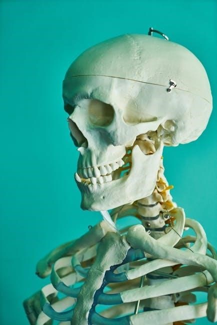
The brain is the control center of the nervous system, governing functions like movement, sensation, and cognition. Understanding its structure is crucial for neuroscience and medicine.
1.1 Overview of Brain Structure
The brain’s structure includes the cerebrum, brainstem, and cerebellum; The cerebrum, divided into lobes, controls higher functions like thought and emotion. The brainstem connects the cerebrum to the spinal cord, regulating vital functions. The cerebellum coordinates movement and balance. Gray matter, containing neurons, and white matter, with myelinated fibers, form the brain’s functional framework, enabling communication and control throughout the body.
1.2 Importance of Studying Brain Anatomy
Studying brain anatomy is essential for understanding nervous system functions, diagnosing disorders, and developing treatments. It aids in neurosurgery, neurology, and psychiatry by mapping brain structures to their roles. This knowledge enhances neuroscience research, improves educational curricula, and supports advancements in neuroimaging and therapeutic interventions, ultimately benefiting human health and cognitive understanding.
Gross Anatomy of the Brain
The brain consists of the cerebrum, brainstem, and cerebellum, each controlling various body functions, sensory processing, and motor activities, forming the structural basis of nervous system operations.
2.1 Cerebrum: Lobes and Functions
The cerebrum is divided into four lobes: frontal, parietal, temporal, and occipital. Each lobe specializes in distinct functions—executive control, sensory processing, memory, and vision—forming the brain’s functional core.
2.2 Brainstem: Structure and Role
The brainstem connects the cerebrum to the spinal cord, comprising the midbrain, pons, and medulla oblongata. It regulates vital functions like heart rate, breathing, and blood pressure, while also managing sensory and motor pathways. Its role is essential for maintaining basic life-sustaining processes and enabling communication between the brain and body.
2.3 Cerebellum: Morphology and Motor Control
The cerebellum, located at the brain’s posterior, has a distinctive folded surface with hemispheres. It specializes in motor coordination, balance, and learning. Damage can impair movement precision and posture, highlighting its vital role in motor function and neuroplasticity.
Microscopic Anatomy of the Brain
The microscopic anatomy reveals neurons and glial cells, with neurons transmitting signals and glial cells providing support. This cellular structure underpins brain function and communication.
3.1 Neurons and Glial Cells
Neurons are specialized cells transmitting signals via synapses, enabling communication. Glial cells, like astrocytes and oligodendrocytes, provide structural and metabolic support. Together, they form the cellular basis of brain function, with neurons handling information transfer and glial cells ensuring a stable environment, crucial for overall nervous system efficiency and adaptability. Their interaction maintains brain health and function effectively.
3.2 Synapses and Neural Circuits
Synapses are junctions where neurons communicate via chemical or electrical signals. Neural circuits, formed by interconnected neurons, process information and enable complex brain functions. Synaptic plasticity, the ability to strengthen or weaken connections, underpins learning and memory. These circuits are essential for sensory processing, motor control, and cognitive tasks, forming the foundation of the brain’s functional operations and adaptability in response to stimuli and experiences.
White and Gray Matter
White matter consists of myelinated axons, enabling rapid signal transmission. Gray matter contains neuron cell bodies and synapses, crucial for processing information and controlling functions. Together, they form the brain’s structural and functional framework, essential for communication and cognition.
4.1 Composition and Function
White matter is composed of myelinated nerve fibers, facilitating rapid neural communication. Gray matter contains neuronal cell bodies and synapses, crucial for processing information. Together, they form the structural basis of brain function, enabling sensory, motor, and cognitive processes. Their composition and organization are essential for maintaining the brain’s operational integrity and facilitating communication across different regions.

4.2 Clinical Relevance
Understanding white and gray matter is crucial for diagnosing neurological disorders. Damage to white matter, often seen in multiple sclerosis, disrupts communication between brain regions. Gray matter abnormalities are linked to conditions like Alzheimer’s disease. Accurate identification of these tissues is vital for MRI interpretations and treatment planning, emphasizing their role in clinical neurology and neurosurgery for improved patient outcomes and targeted therapies.

Cranial Nerves
Cranial nerves connect the brain to various body parts, controlling functions like movement, sensation, and bodily reactions. They are essential for maintaining vital physiological responses and motor control.
5.1 Classification and Functions
Cranial nerves are classified into twelve pairs, each serving specific roles. They regulate sensory input, motor functions, and autonomic processes. Functions range from controlling eye movements to managing digestion and heart rate, ensuring the body operates harmoniously under the brain’s command.
5.2 Clinical Significance
Cranial nerves’ dysfunction can lead to severe conditions like facial paralysis, vision loss, or swallowing difficulties. Clinical assessment of these nerves aids in diagnosing disorders such as strokes or brain tumors, emphasizing their critical role in neurological evaluations and patient care.

Blood Supply to the Brain
The brain receives blood through two main routes: internal carotid arteries and vertebral arteries, forming a network crucial for oxygen and nutrient delivery. Blockages here can cause strokes.
6.1 Arterial Supply
The brain’s arterial supply primarily originates from the internal carotid arteries and vertebral arteries. These vessels branch into a complex network, including the circle of Willis, ensuring robust blood flow to cerebral tissues. This dual supply enhances redundancy, protecting against ischemic events. Proper arterial function is vital for maintaining cognitive and motor capabilities.
6.2 Venous Drainage
Brain venous drainage relies on dural sinuses and cortical veins. These structures collect deoxygenated blood and return it to the heart via jugular veins. The superior and inferior sagittal sinuses are key components, ensuring efficient blood circulation. Proper venous drainage is essential for brain health to prevent conditions like venous thrombosis.
Functional Anatomy of Brain Regions
Brain regions like the frontal, temporal, parietal, and occipital lobes specialize in distinct functions, from executive tasks to sensory processing, enabling integrated brain activity.
7.1 Frontal Lobe: Executive Functions
The frontal lobe is responsible for executive functions, including decision-making, planning, and problem-solving. It regulates emotions, motivation, and impulse control, serving as the center for higher-order thinking and goal-oriented behavior.
7.2 Temporal Lobe: Memory and Hearing
The temporal lobe plays a vital role in auditory processing and memory formation. It houses the hippocampus, essential for converting short-term memories into long-term ones. Damage to this lobe can impair hearing and memory recall, highlighting its significance in cognitive and sensory functions.
7.3 Parietal Lobe: Sensory Processing
The parietal lobe processes sensory information, including touch, temperature, and spatial awareness. It aids in navigating environments and manipulating objects. Damage can lead to impaired spatial perception and sensory deficits, emphasizing its role in integrating sensory data for motor responses and environmental interaction.
7.4 Occipital Lobe: Vision
The occipital lobe is primarily responsible for processing visual information. Located at the back of the brain, it interprets signals from the eyes, enabling us to recognize shapes, colors, and patterns. Damage to this area can result in vision loss or impaired visual perception, highlighting its critical role in interpreting sensory data for meaningful visual experiences.

Diencephalon
The diencephalon includes the thalamus, hypothalamus, and epithalamus, playing a central role in sensory processing, hormone regulation, and maintaining homeostasis. It acts as a relay station for sensory information and controls vital functions like hunger, sleep, and body temperature, ensuring proper physiological balance.
8.1 Thalamus: Relay Station
The thalamus acts as a critical relay station, processing and directing sensory information to the cortex. It refines signals from sensory organs, ensuring precise transmission. This structure is essential for consciousness, regulating the flow of information between the brain and body, and facilitating higher cognitive functions by filtering and prioritizing sensory data effectively.
8.2 Hypothalamus: Hormonal Regulation
The hypothalamus is a vital regulator of hormonal balance, controlling the pituitary gland and influencing the endocrine system. It produces hormones like oxytocin and vasopressin, managing body temperature, hunger, and thirst. Additionally, it coordinates the body’s response to stress, ensuring homeostasis and integrating neural and hormonal signals to maintain overall physiological stability and health.

Brain Ventricles and Cerebrospinal Fluid
The brain ventricles produce cerebrospinal fluid (CSF), which cushions the brain, regulates pressure, and circulates nutrients and waste, ensuring neural protection and homeostasis.
9.1 Structure and Function
The brain ventricles are interconnected chambers within the brain, producing cerebrospinal fluid (CSF). The lateral ventricles, located in the cerebral hemispheres, and the third and fourth ventricles form a system that circulates CSF, maintaining intracranial pressure, cushioning the brain, and removing waste products, essential for central nervous system health and function.
9.2 Clinical Implications
Abnormalities in brain ventricles, such as hydrocephalus, can lead to increased intracranial pressure, cognitive deficits, and motor dysfunction. Accurate imaging and understanding of ventricular anatomy are critical for diagnosing conditions like hydrocephalus or hemorrhage. Proper CSF circulation is vital for brain health, and disruptions can result in severe neurological complications, emphasizing the importance of precise neurosurgical interventions.
Neuroanatomical Pathways
Neuroanatomical pathways connect brain regions, enabling communication. They include motor and sensory routes, crucial for voluntary actions and perception, forming the structural basis of neural function and behavior.
10.1 Motor Pathways
Motor pathways are neural networks enabling voluntary movement. The corticospinal tract, originating in the cerebral cortex, transmits signals to the spinal cord, controlling limb movements. Other pathways, like the corticobulbar tract, regulate facial and neck muscles. Damage to these pathways can result in motor impairments, highlighting their critical role in movement coordination and control.
10.2 Sensory Pathways
Sensory pathways transmit information from sensory receptors to the brain. The dorsal column pathway conveys touch and vibration, while the spinothalamic tract handles pain and temperature. These pathways ascend through the spinal cord and brainstem to the thalamus, then to the cerebral cortex, enabling perception and interpretation of sensory stimuli, crucial for environmental interaction and survival.
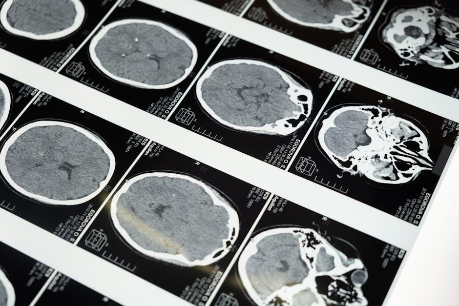
Imaging Techniques in Brain Anatomy
Advanced imaging techniques like MRI and CT scans provide detailed visualization of brain structures. These tools are essential for diagnosing abnormalities and understanding brain anatomy in research and clinical settings.
11.1 MRI and CT Scan
Magnetic Resonance Imaging (MRI) and Computed Tomography (CT) scans are pivotal in visualizing brain anatomy. MRI provides detailed images of soft tissues, while CT scans offer quick assessments of structural abnormalities. Both techniques are essential for diagnosing neurological conditions and understanding brain morphology in clinical and research settings.
11.2 Functional Neuroimaging
Functional neuroimaging techniques like fMRI, PET, and EEG map brain activity and connectivity. They reveal how different regions interact during tasks, aiding in understanding cognition, behavior, and neurological disorders. These tools are invaluable for both research and clinical applications, providing insights into the dynamic processes of the brain.
Clinical Applications of Brain Anatomy
Brain anatomy knowledge is vital for neurosurgery and neurology, enabling precise tumor removals, stroke diagnostics, and treatment planning. It guides interventions, improving patient outcomes and surgical accuracy.
12.1 Neurosurgery
Brain anatomy is critical in neurosurgery, guiding tumor resections, aneurysm repairs, and epilepsy surgeries. Detailed knowledge of brain structures ensures precise interventions, minimizing damage to vital areas like motor and language centers.
12.2 Neurology
Brain anatomy is fundamental in neurology for diagnosing disorders like strokes, tumors, and dementia. Understanding gray and white matter, cranial nerves, and blood supply aids in localizing lesions and treating conditions affecting motor, sensory, and cognitive functions.